Jena research team develops novel microscope for cancer diagnostics

14. Dezember 2020
Researchers from Jena, together with European partners from research, medicine and industry, work on a novel microscopy technology.
It should help to track down the cellular origins of cancer diseases and decisively advance precision medicine. With this goal in mind, the team from the Leibniz Institute of Photonic Technology (Leibniz IPHT), Jena University Hospital (UKJ) and laser system manufacturer Active Fiber Systems launches the transnational transdisciplinary research project CRIMSON (Coherent Raman Imaging for the Molecular Study of the Origin of Diseases) in December 2020.
Together with other leading research institutions and companies from Italy, the UK and France, they are developing a biophotonic imaging device based on next-generation coherent Raman microscopy for biomedical research. It combines advanced laser techniques with data analysis through artificial intelligence. The European Commission is funding the project with more than €5 million over 42 months.
Innovative microscopy technology for imaging inside the body
“Our goal is to bring an innovative, label-free microscopy technology to the market and to the clinic that will make it possible to detect changes in cells based on a molecular fingerprint,” explains Prof. Jürgen Popp, scientific director of Leibniz IPHT. In perspective, the microscopic method will also be used for endoscopic imaging inside the body and enable rapid, high-precision tissue diagnostics. “This innovative technology will enable us to better understand the interaction between cancer cells in the head and neck region and the cells of the immune system,” adds Prof. Orlando Guntinas-Lichius, director of the Department of Otolaryngology at the UKJ. For the planned device, the Jena-based company Active Fiber Systems (AFS) is developing a new type of compact fiber laser, which will then enable the researched microscopic method to be used directly in the clinic in the future. “This light source will be used for various imaging procedures,” said AFS development manager Tino Eidam.
The breakthrough microscope will provide three-dimensional molecular images of subcellular compartments in living cells and organoids, enabling rapid tissue classification with unprecedented biomolecular sensitivity. The high imaging speed allows observation of intra- and intercellular dynamic changes at high frame rates. In addition to the microscope, the research teams are also developing an innovative endoscope for imaging inside the body. To simulate future in vivo studies, it will initially be used to examine thick tissue samples ex vivo.
Multidisciplinary team from leading international research institutions
A multidisciplinary team of leading international research institutions and companies make up the project’s consortium, which is coordinated by the Politecnico di Milano. Three research centers with long-standing expertise in photonics, spectroscopy and nonlinear microscopy will develop the microscopy/endoscopy technology: In addition to the Jena-based Leibniz Institute of Photonic Technology, these are the Politecnico di Milano (Italy) and the Centre National de la Recherche Scientifique (France). Biomedical partners, in addition to Jena University Hospital, are the Instituto Nazionale Tumori (Italy) and the Institut National de la Santé et de la Recherche Médicale (France). They will validate the imaging system with regard to open biological questions in cancer research. These are paradigmatic for the complexity and heterogeneity of cellular diseases.
Active Fiber Systems GmbH and three other innovative SMEs (Lightcore Technologies, France; Cambridge Raman Imaging Limited, UK; 3rdPlace S.r.l., Italy) — including a biomedical device manufacturer — will commercialize the innovation. In doing so, they will create a competitive advantage in the European market for microscopes and R&D tools in the field of biophotonics. The results have the potential to improve patients’ quality of life and reduce public health costs.
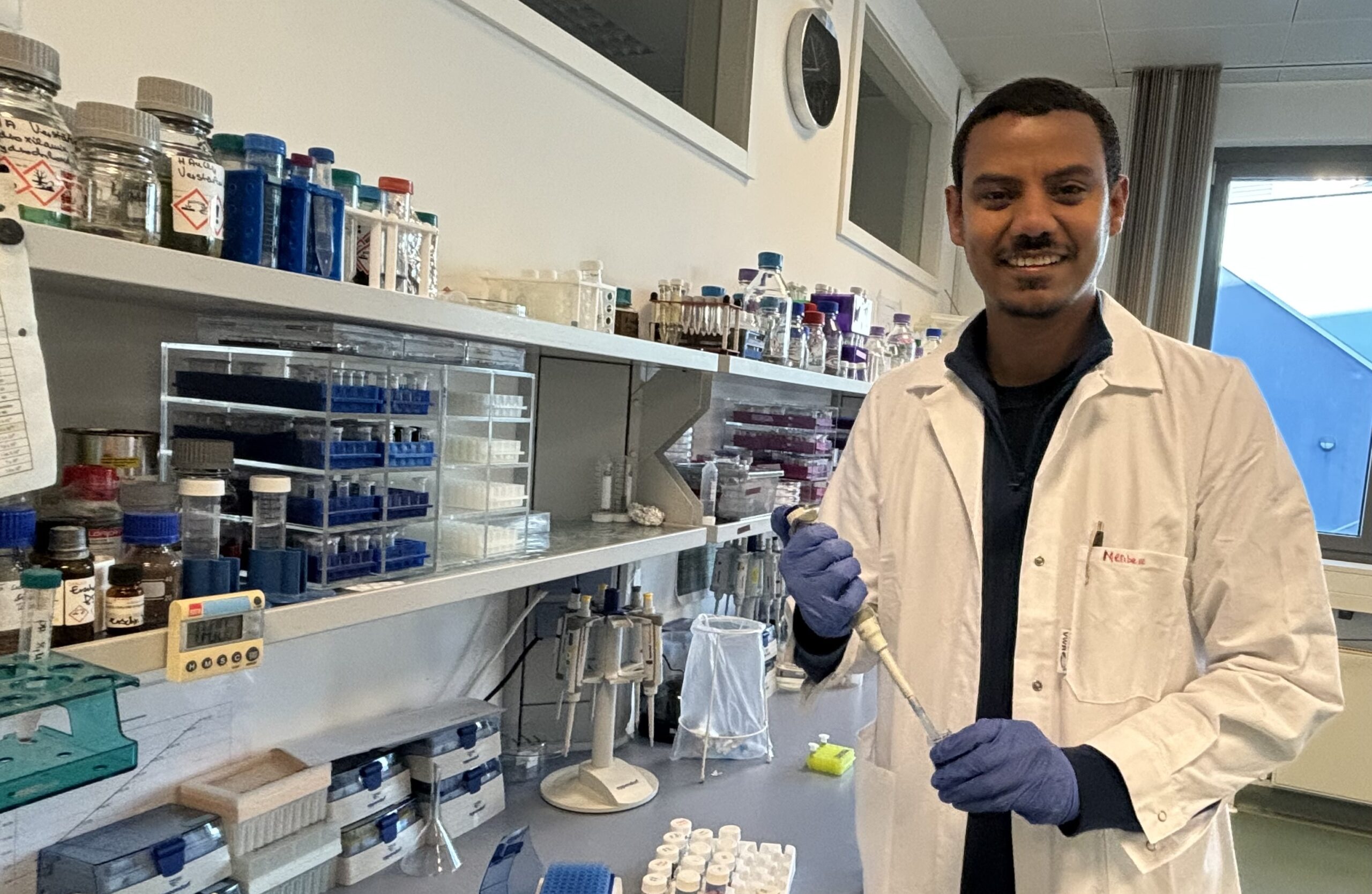
For rapid cancer diagnostics: CRIMSON partner Tobias Meyer-Zedler from Leibniz-IPHT is researching laser-based, multimodal imaging techniques to visualize biomolecules in cells, tissues and organs. // Photo: Sven Döring/ Leibniz-IPHT
Contact

Related News
Third party cookies & scripts
This site uses cookies. For optimal performance, smooth social media and promotional use, it is recommended that you agree to third party cookies and scripts. This may involve sharing information about your use of the third-party social media, advertising and analytics website.
For more information, see privacy policy and imprint.
Which cookies & scripts and the associated processing of your personal data do you agree with?
You can change your preferences anytime by visiting privacy policy.