- Home
- Research
- Spectroscopy and Imaging
Scientific Profile
The research department focuses on the research, development and implementation of innovative optical/photonic methods and tools for multiscale spectroscopy and multimodal imaging methods together with particle- and chip-based molecular point-of-care concepts for analytics, diagnostics and therapy in the fields of medicine, life and environmental sciences, quality and process analytics and pharmacy. The implementation of novel fibre concepts for spectroscopy and imaging as well as fibre-based sensor technology, strategies for spectroscopic high-throughput measurements and field-resolved optical precision measurement methods open up new analytical fields of application. The use of surface-enhancing Raman and IR detection methods to increase the sensitivity of spectroscopic methods round off the department's research profile. In general, the research department pursues the goal of covering the entire analytical measurement chain, whereby sample preparation and processing methods that can be used decentrally and independently of the laboratory are investigated and implemented in the overall workflow. Statistical, chemometric and image analysis methods as well as machine learning are researched and used to analyse the data obtained.

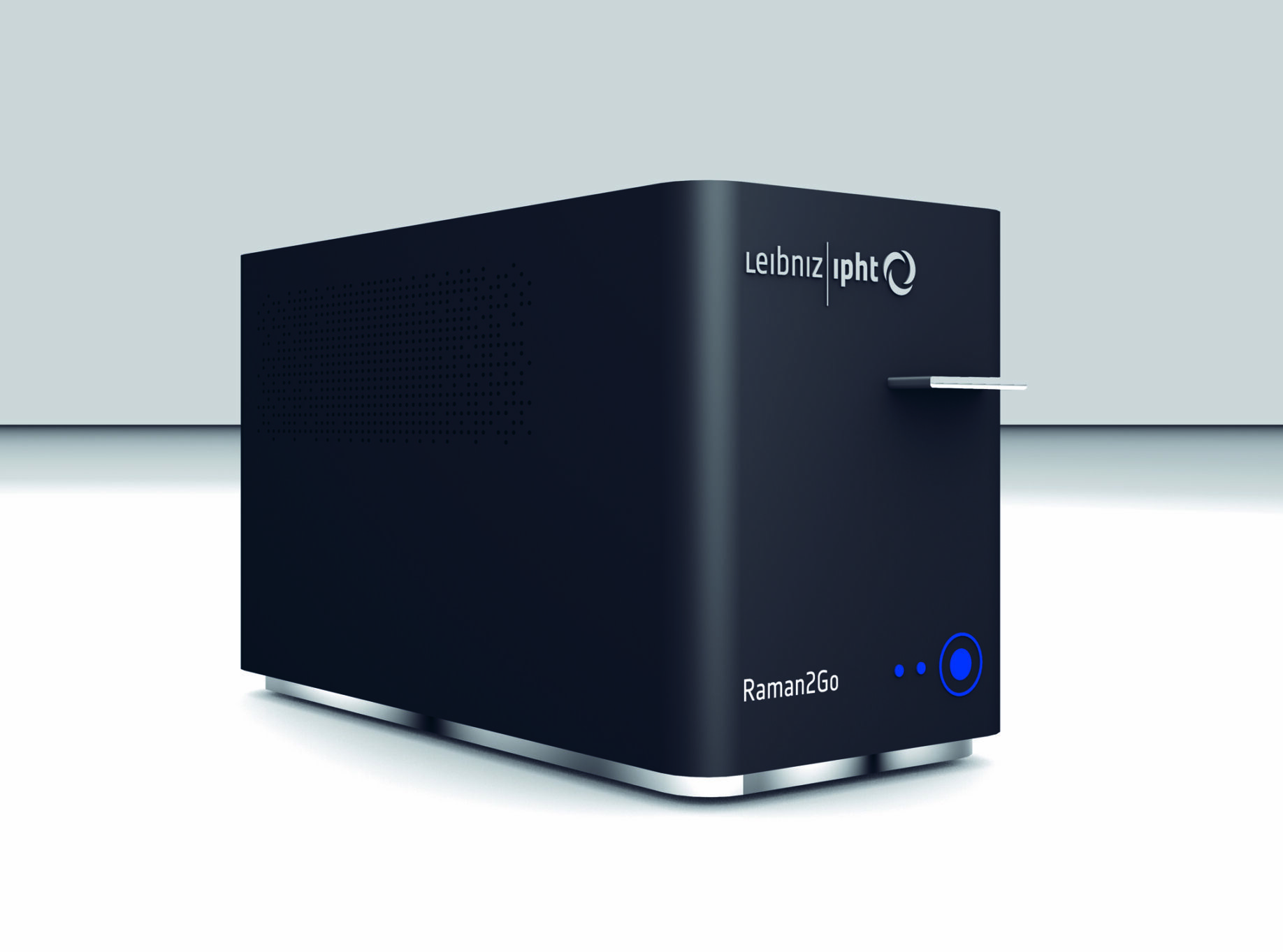
Design of a miniaturized Raman system

Compact multimodal nonlinear microscope for intraoperative frozen section diagnostics
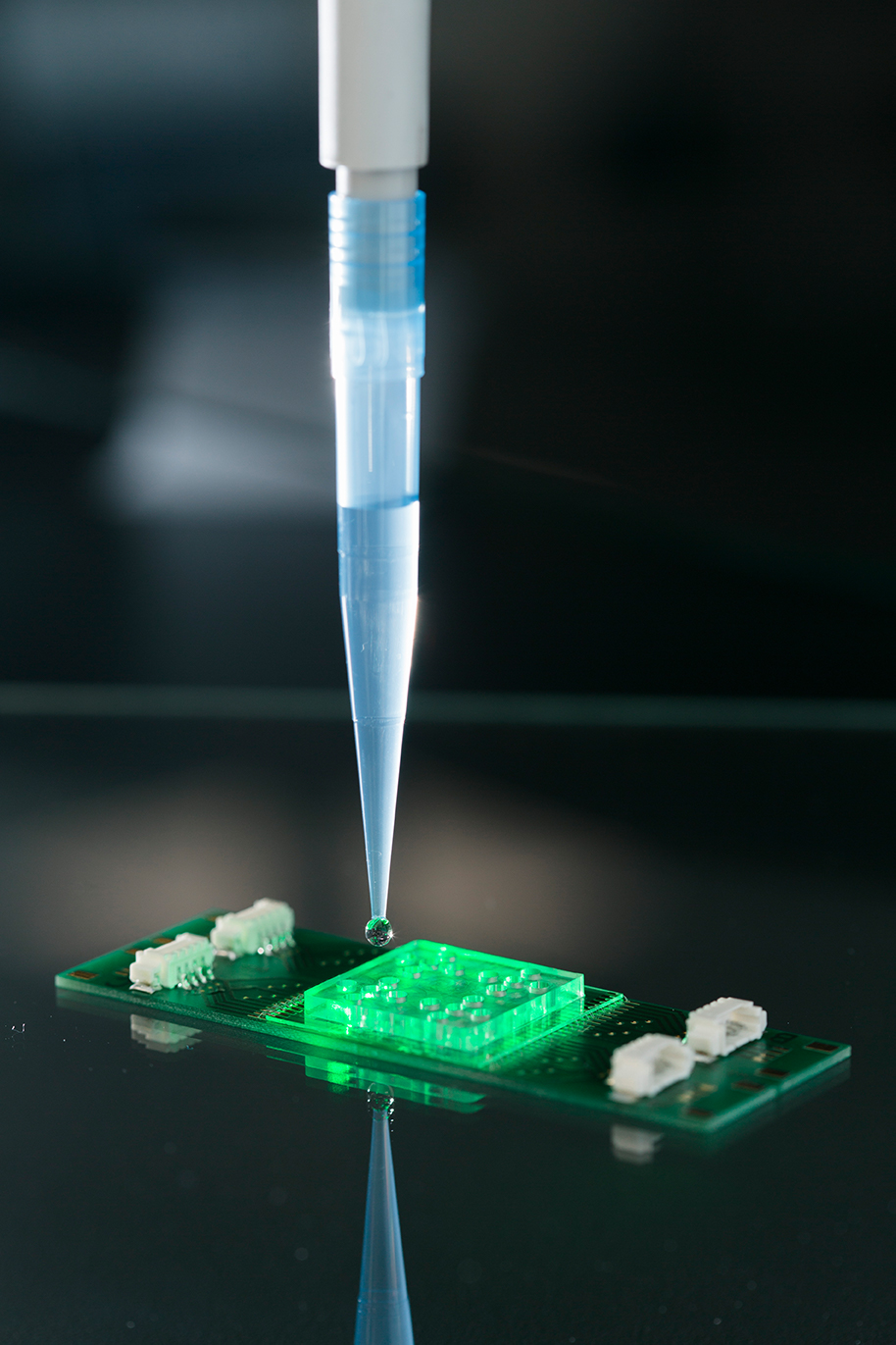
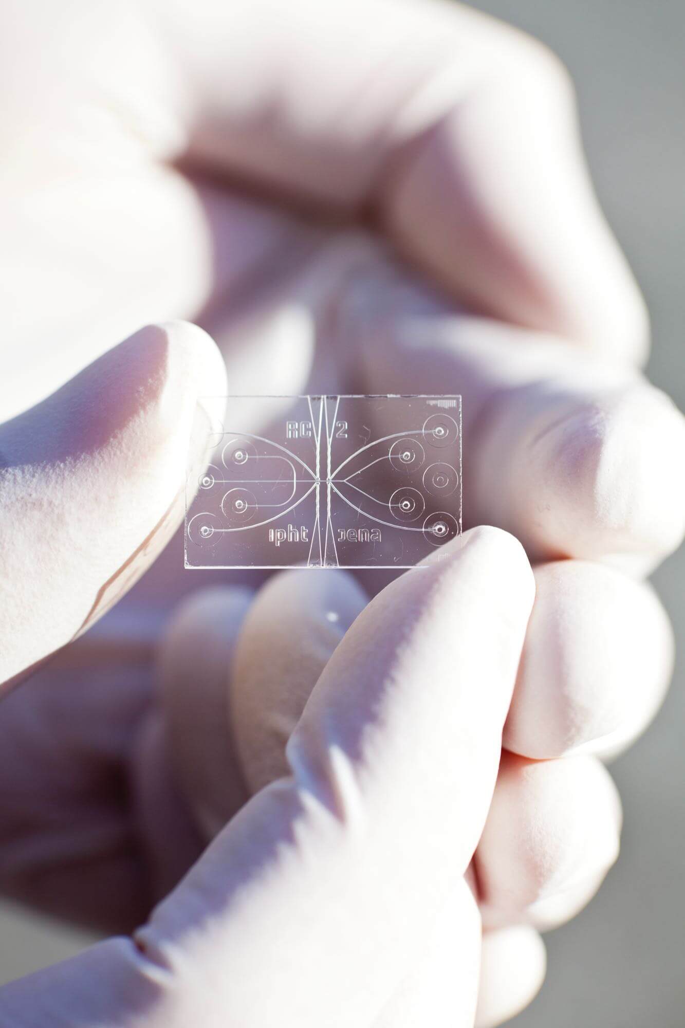
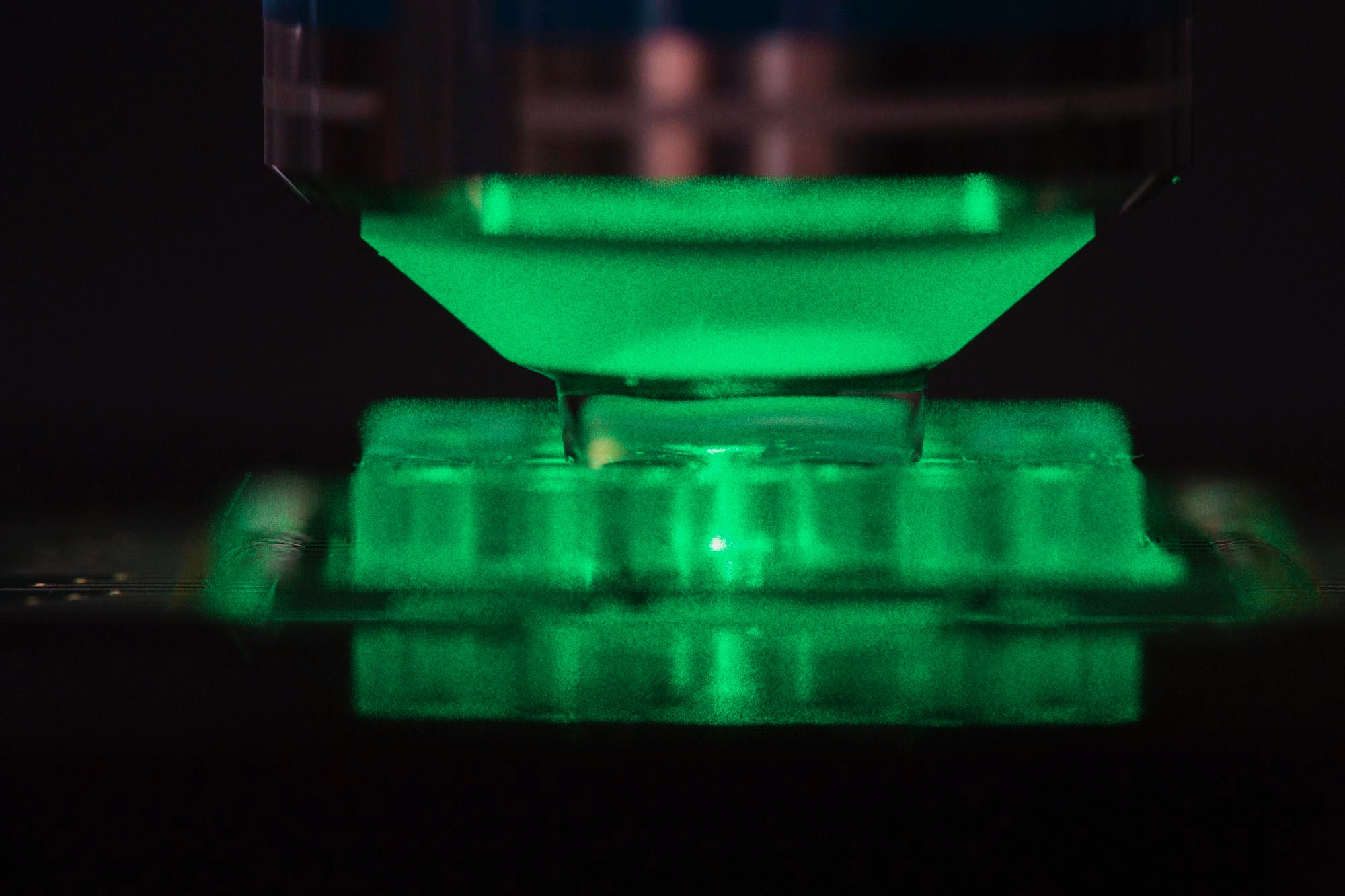
Dielectrophoretic chip with 20 cavities for Raman-based pathogen diagnosis and resistance determination
Research Topics
- Methodical development of molecular and functional contrast mechanisms
- Raman / multimodal instrumentation for biomedical questions; implementation of imaging techniques in compact systems; mixed- and augmented reality;
- Research and realization of miniaturized low-cost Raman systems
- Raman spectroscopic characterization and identification of pathogens and their antibiotic resistance (RamanBioAssay)
- Implementation of spectral sensors, microfluidics, optical fibers, chip- and nanoparticle-based concepts in compact miniaturized setups;
- Research on on-site and laboratory independent sample preparation and treatment strategies as essential tools for the success of new detection systems and methods to cover the entire analysis chain;
- Research activities on surface enhanced detection techniques to increase the sensitivity of spectroscopic methods for life science applications;
- Highly sensitive Raman gas sensors to investigate environmental issues;
- Investigation of novel hollow core fibers for highly sensitive Raman sensor applications ;
- Pattern recognition, multivariate and hyperspectral data analysis (data visualization, processing); device control.
The search for new molecular and functional contrast mechanisms, their implementation in innovative spectral-optical methods and the instrumentation based on them are a central challenge. Here, two complementary approaches are pursued: While a top-down approach based on physical measurement methods can be used to increase specificity and sensitivity, the research, development, and integration of innovative markers and labels represents a bottom-up approach to chemical contrast.
The research, development and implementation of innovative technologies and processes are particularly aimed for:
- improving disease diagnosis (qualitatively and quantitatively);
- providing in-depth insights into the dynamics of life processes;
- opening up new fields of application for optical technologies for biomedical, environmental and life science cooperation partners.
Areas of application
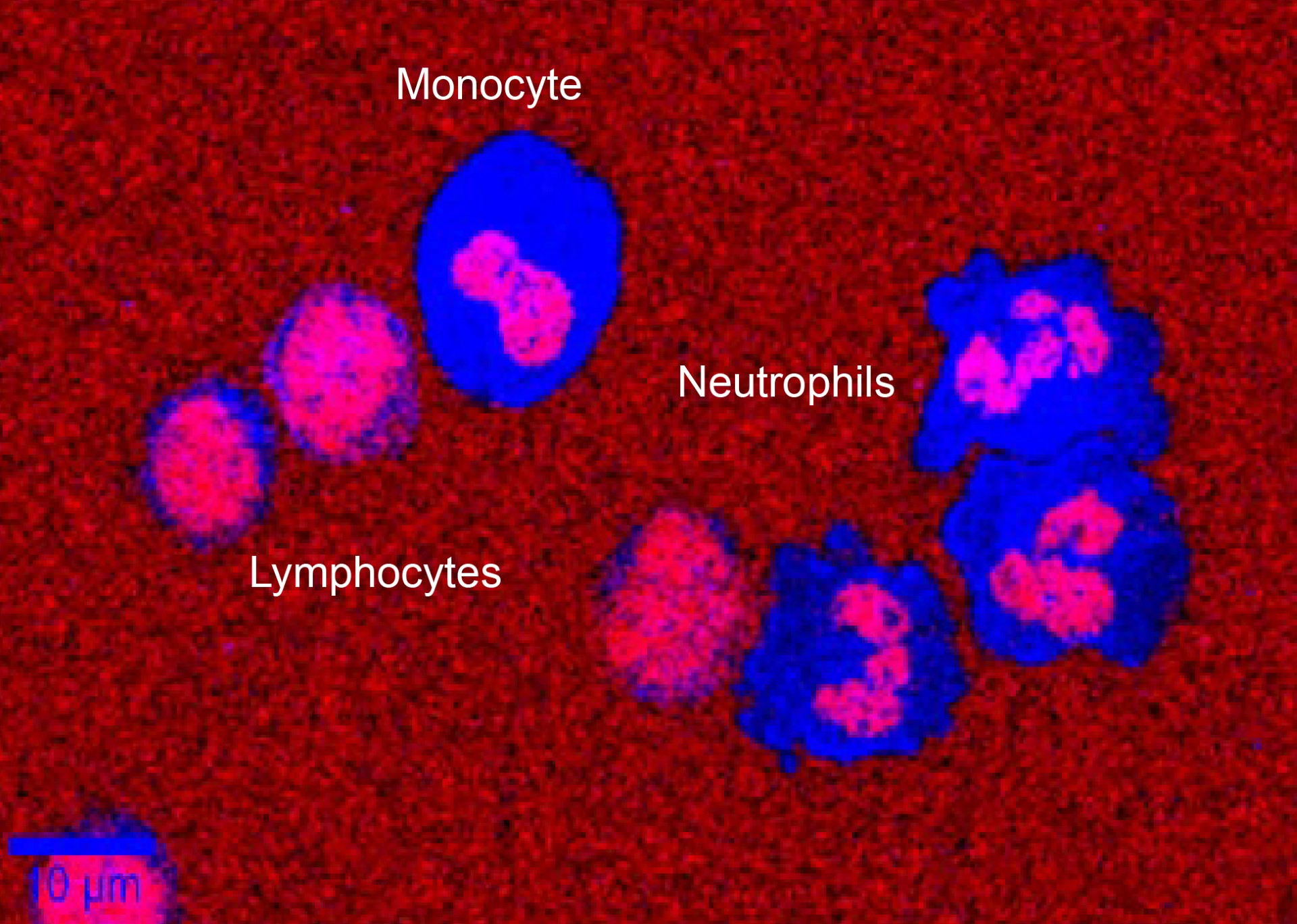
- Microbial photonic diagnostics (pathogen / host / companion)
- infection diagnostics
- Spectral histopathology (molecular spectroscopy, high-resolution and hyperspectral imaging)
- Medical, environmental, pharmaceutical, food and process analytics