- Home
- Research
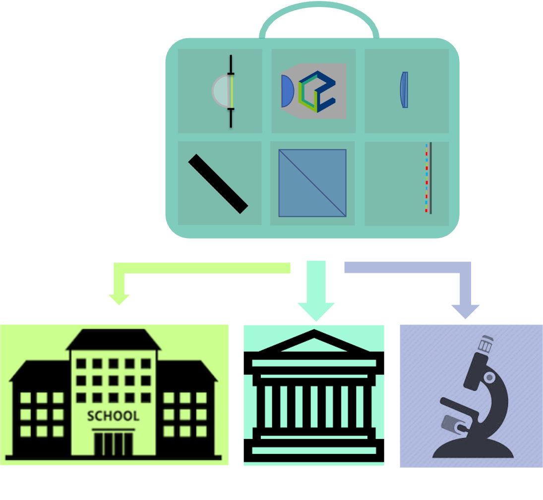
- Microscopy
Scientific Profile
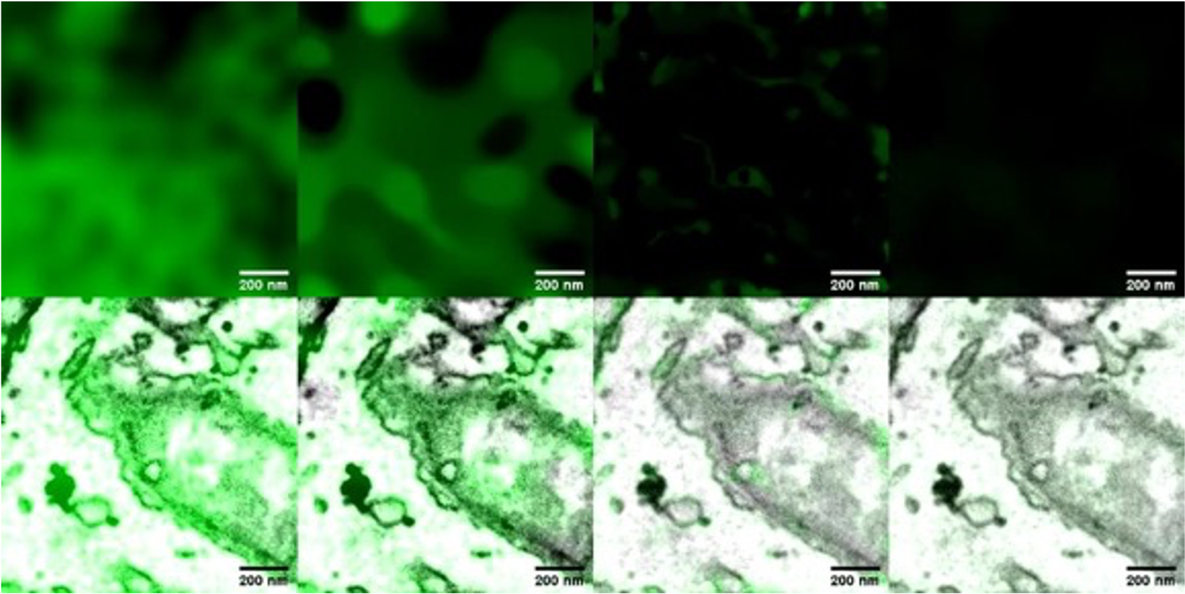
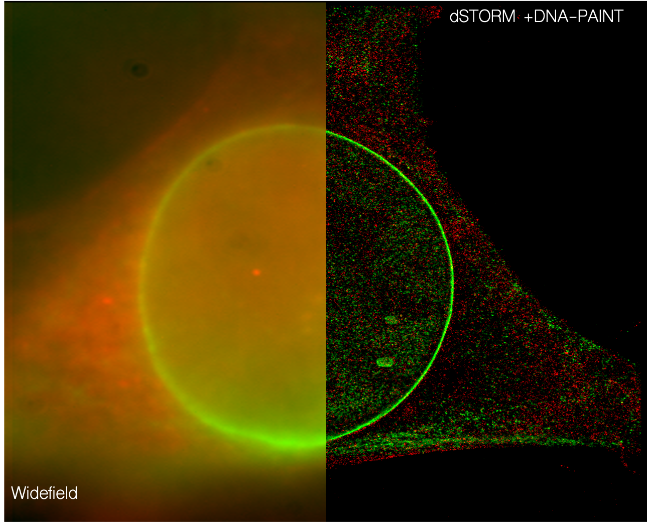
With the help of modern microscopic methods, biomedical mechanisms can be made visible in the submicrometer range. The development of new methods and instruments for light microscopy and the advancement of known techniques are the main focus of our research department. We make use of the fact that light is ideally suited as a non-destructive sensor to develop methods, characterize materials and investigate life processes. Close cooperation with biological and medical research groups enables individual and application-oriented optimization with the aim of better understanding biomedical processes. This is accompanied by the development of computer-aided image reconstruction methods based on programs such as Matlab, Python and GPU (graphics processing unit) programming in CUDA (Compute Unified Device Architecture). One goal is, for example, the dynamic representation of receptor functions or polymicrobial infections in real time. We use modern techniques, such as microscopy with spatial resolution beyond the Abbe diffraction limit, and are working on the development of inexpensive but powerful microscopes for education and research using modern 3D printing techniques (useetoo.org), as well as algorithms for image analysis.
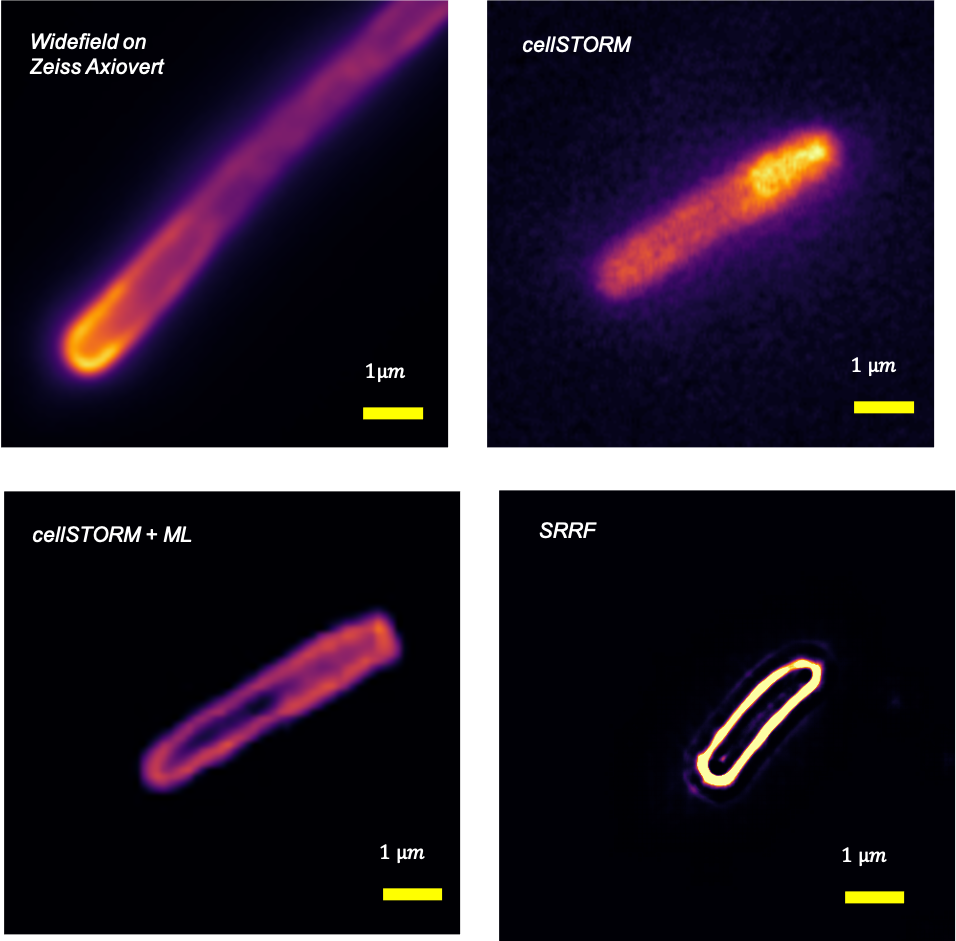
Cellphone-Based Super-resolution Imaging: „cellSTORM“
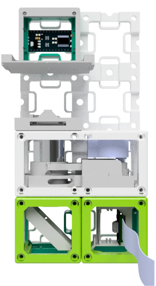
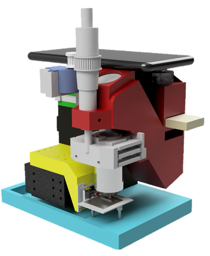
Open-Source Modular Microscopy: „UC2“
Open Education in Photonics and Research
Research Topics
- Fluorescence super-resolution microscopy
- Alternative microscopic methods
- Improvement of image reconstruction algorithms
- Highly sensitive detection of minimal losses in optical materials and coatings
Areas of application
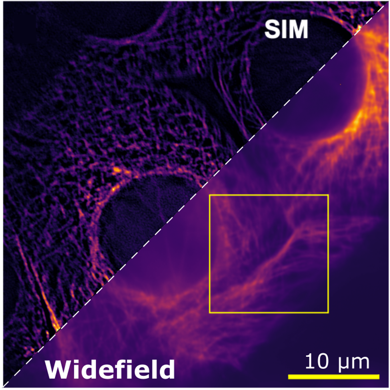
- Use of optical microscopy, e.g. modern high-resolution fluorescence microscopy (SIM, dSTORM, etc.) for biomedical questions
- Hyperspectral Raman imaging for medical research
- Solution of inverse problems in microscopy (e.g. image deconvolution and reconstruction of 3D images) with neural networks
- Inexpensive microscopes for research and education using 3D printing and smartphones
- Material characterization in an industrial environment