- Home
- Research
- Spectroscopy and Imaging
- Work-Groups
- Applied Biospectroscopy and Bioassays
Applied Biospectroscopy and Bioassays
A major focus of the Applied Biospectroscopy and Bioassays group is to advance and standardize the application of optical detection methods with regard to important fields of application in the life sciences and medicine and, last but not least, to make them more user-friendly. Amongst other detection methods Raman spectroscopy is applied, as it allows retrieving highly specific information regarding the molecular composition of various biological samples. For example, the characterization of cellular phenotypes is realized, but also in-vitro studies with active substances are carried out. As Raman spectroscopy is generally non-destructive, the use of complementary methods such as PCR (polymerase chain reaction) or NGS (next generation sequencing) is easy and straightforward, so that comprehensive information on the samples can be obtained.
Through the concurrent development of customized sample preparation strategies for different issues, the potential of the detection methods used is fully exploited. The implementation of such strategies in the process or analysis chain makes it possible to address samples of complex composition and to work with very small volumes. By automating process steps and introducing standards and references, assays can be established with a high degree of reliability.
For developing novel sample preparation methods, the working group is researching different approaches for surface modification. The aim here is to generate functional interfaces that allow, for example, the targeted enrichment of analytes or cells. By using surface-sensitive methods, not only can the quality of the functionalization be evaluated, but interactions between capture molecules or recognition elements and analytes can also be investigated. Accordingly, these activities play a vital role in the development of innovative sample preparation strategies.
Contact

Principle of antibiotic susceptibility testing using Raman spectroscopy. The bacteria are enriched in the center of the chip by dielectrophoresis (a). Subsequently, their Raman spectrum is detected (b). The Raman spectrum reveals whether the growth of a bacterium is inhibited by a specific antibiotic (c).
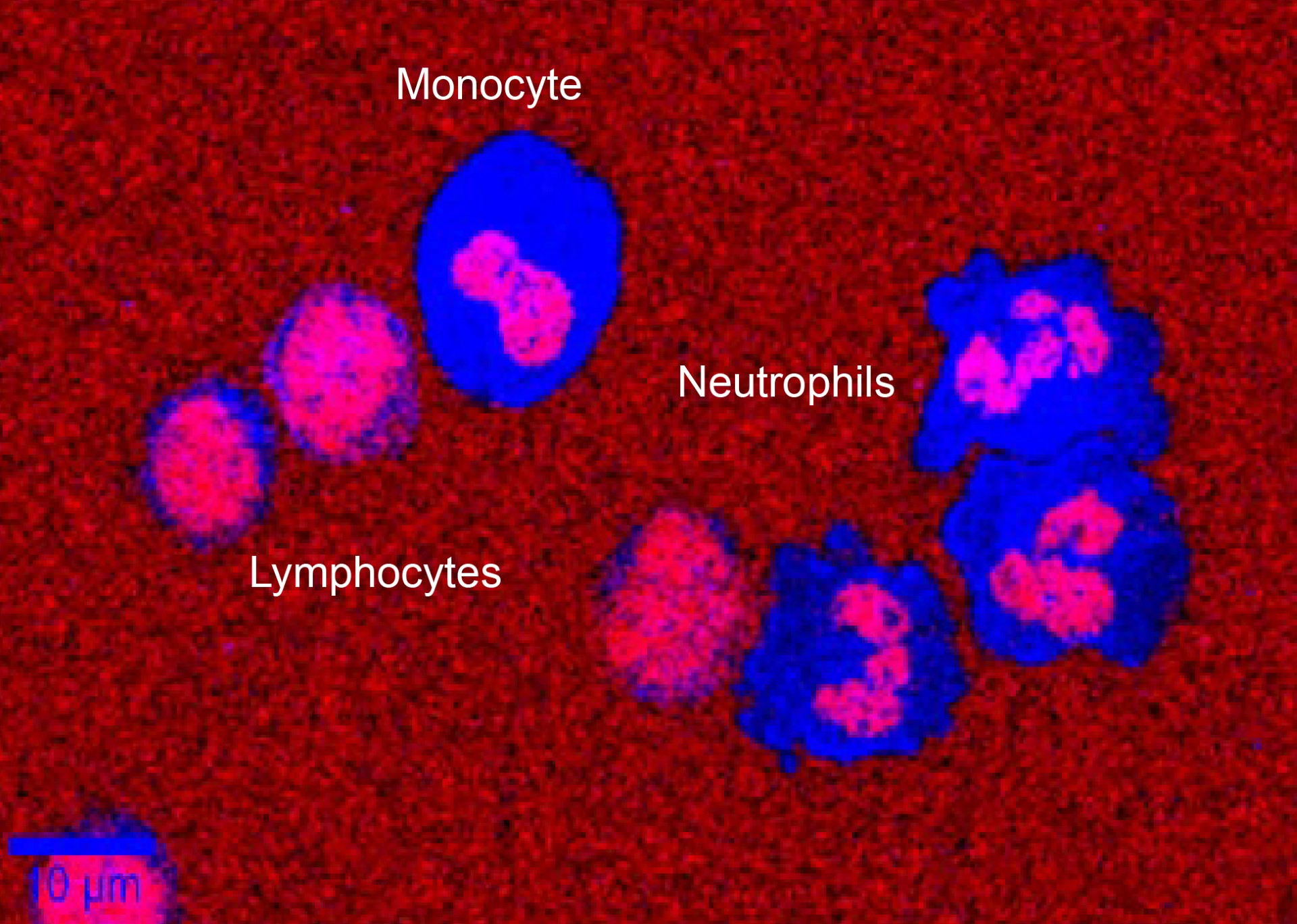
The potential of Raman spectroscopy as a diagnostic method is being researched in clinical studies. The image shows a false color map of different types of white blood cells (leukocytes), which was generated using characteristic Raman bands. By determining the number and ratio of certain types of leukocytes, conclusions can be drawn about the patient's state of health.

Magnetic particles functionalized with a fluorescence-labelled ACE2 receptor (red) on the surface of which fluorescent SARS-CoV-2 pseudoviruses (green) were captured. Collaboration with Dr. Ziliang Zhao from the Department of Biophysical Imaging as well as Dr. Stefanie Reuter and Prof. Dr. Mrowka from Jena University Hospital.

Droplet-based assays enable rapid and sensitive detection of bacteria. Different phenotypes can be differentiated using chromogenic or fluorogenic substrates. The image shows E. coli cells that were isolated using a droplet assay. A color change occurs in the droplets with E. coli (reddish color) because the bacteria contain an enzyme that catalyzes the conversion of the dye.
Forschungsthemen
- spectroscopic investigation of cellular phenotypes, i.e. in-vitro interaction studies
- Development of Raman-spectroscopic diagnostic assays for cells
- Chip- and particle-based sample preparation strategies
- Research into strategies for the enrichment of intact cells
- Research into methods for surface functionalization and characterization
Areas of application

- Identification of novel targets for the diagnosis of infectious diseases
- Enrichment of viruses with magnetic beads for subsequent Raman spectroscopic Identification
- Reversible binding of bacteria to magnetic beads
- Biomarker detection with interference-enhanced Raman spectroscopy
- Phenotypical antibiotic resistance testing
On the image: A false color map of different types of white blood cells (leukocytes), which was generated using characteristic Raman bands