- Home
- Research
- Quantum Systems
- Research results
- Biomagnetic Imaging with Optically Pumped Magnetometers
Biomagnetic Imaging with Optically Pumped Magnetometers

08.11.2019
The magnetometry research group is investigating new optically pumped magnetometers that enable biomagnetic imaging in external magnetic fields as strong as the Earth’s field on various scales: the large-area recording of human biomagnetic signals and the microscopic imaging of the fields of small animals.
By V. Schultze // R. IJsselsteijn // R. Stolz // F. Wittkämper // G. Oelsner // R. Eichardt // J. Haueisen // G. Oelsner // C. B. Schmidt // U. Graichen
The measuring principle developed at Leibniz IPHT for new optically pumped magnetometers (OPMs) offers the unique possibility of achieving very high magnetic field resolutions when measuring in the Earth’s magnetic field [1]. This paves the way not only for applications in geomagnetic surveying [2] but for biomagnetic imaging without complex and expensive magnetic shielding as well. Current developments aim at the detection of magnetic fields of living organisms on different size scales. In this contribution, two instruments will be examined in more detail: This concerns the larger-scale imaging of a large variety of magnetic signals generated by the human body on a size scale consisting of centimeters. In medicine, the focus of interest is the recording of signals of human brain activity in adults (magnetoencephalogram (MEG)) and cardiac activity in adults (magnetocardiogram (MKG)) and unborn children (fetal MKG (fMKG)). For these types of measurements, modules for one OPM channel each are under development which have a task-oriented configuration and are arranged in arrays. One example of such a module is shown in Figure 1. The actual magnetometer consists of two directly interconnected areas of a common cesium vapor cell, which are irradiated at a distance of 5 mm by two circularly polarized beams of opposite helicity. This enables operation in LSD-Mz mode [1] (see also the 2017 Annual Report). Two magnetometers, which are 40 mm apart but integrated in a common silicon substrate and thus well balanced, enable the suppression of interfering signals, for example by the differential or gradiometric measurement of biomagnetic signals.
The power to configure arrays of such modules for the desired measurement was calculated in simulations exemplarily for the detection of neuronal sources in the human brain using the boundary element method [3]. This makes it possible to determine the dependence of the attainable information on brain activity based on the orientation, distance, and location of the OPM modules. One example in Figure 2 shows the qualitative distribution of the estimated measurement information for 128 standardized sensor positions (corresponding to 10/10-EEG) for a source configuration that extends across the entire cortex (gray dots). Figure 3 shows the changes in information as a function of the sensitivity direction of the OPM (rotation angle) and the index of the sensor position, in which the position has a significantly greater influence on the information content compared to the direction. The results will soon be validated by phantom measurements.
To measure the distribution of magnetic signals from small animals in the millimeter range (e.g., from the heart of mice), an imaging method is being developed that uses a novel magnetic field camera and LSD-Mz mode to read out the optical magnetometer. In contrast to conventional OPMs, a total of 52 magnetometers at different locations of the cell, as well as four reference sensors, are to be implemented in a single, expanded cesium cell of more than 30 mm x 30 mm. The structure of the magnetic field camera and beam guidance with 56 individual laser beams are shown in Figure 4. An adaptation of the calculations mentioned above shows that with this “OPM screen” lateral resolutions of around 6 mm at a distance of about 7 mm from the measuring head housing, as well as realistic signal-to-noise ratios, are possible.
The microsystem structure of the magnetometers developed at Leibniz IPHT (see Fig. 1), which was implemented together with the micro and nanotechnologies group, not only offers the possibility of integrating differently configured arrangements of OPM measuring cells into silicon substrates but can also be combined with microfluidic components such as those being developed by IPHT’s nanobiophotonics research department. This combination of different technological possibilities is used in another project, in which a completely different biomagnetic measurement is to be carried out: the quantification of magnetotactic bacteria, microorganisms which orient themselves in the Earth’s field using their own magnetosomes and which make up a noticeable proportion of the Earth’s microbiological biomass.
Funded by: EU, DFG, BMBF, Freistaat Thüringen
OPM module for one measuring channel: a) Integrated arrangement of two magnetometers with a common cesium reservoir in the middle b) Encapsulation of the module with optics for processing the pump light (in the neck) and the B1 coils around the two magnetometers.
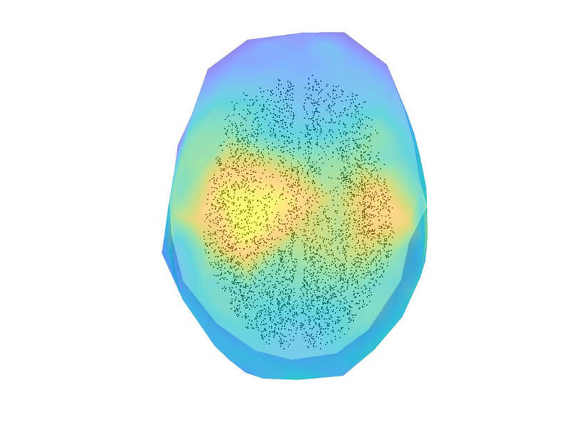
Examined sensor positions on the human head (view from above with the model facing north). (V. Schultze et al., Sensors 17 (2017), 561)
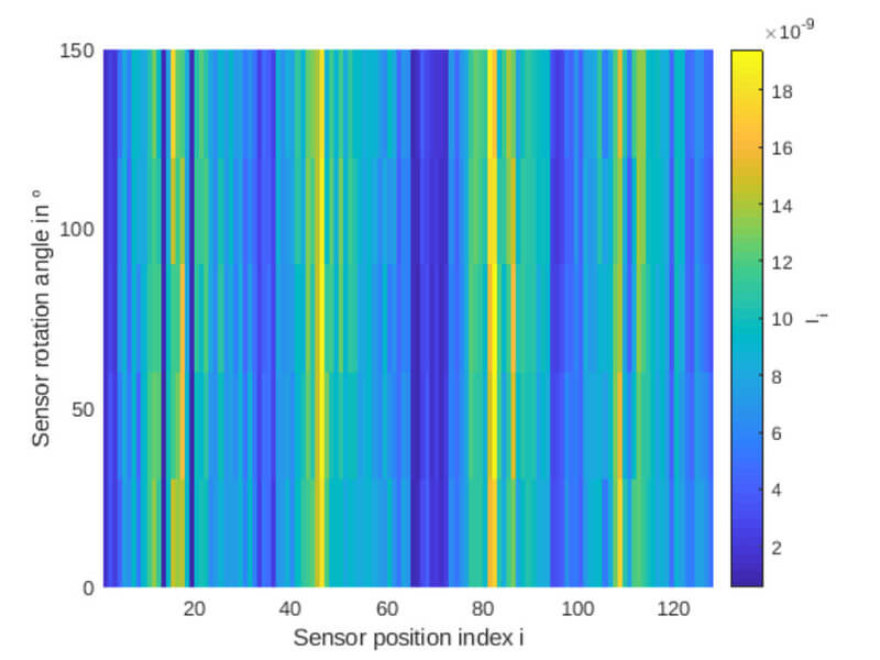
Total information that can be obtained from the 128 different positions in Fig. 2 and different angles to the human head with the OPM modules in Fig. 1 [3]. (V. Schultze et al., Sensors 17 (2017), 561)

Principle sketch of the OPM magnetic field camera. (V. Schultze et al., Sensors 17 (2017), 561)