- Home
- Research
- Microscopy
- Research results
- Enhanced microscopic phase-contrast images using “smart” algorithms implemented on a smartphone
Enhanced microscopic phase-contrast images using “smart” algorithms implemented on a smartphone

23.04.2018
A recent study suggests that about 2.3 billion smartphones are in active use. Most of them are equipped with high-quality cameras and powerful CPUs or even GPUs. This opens new possibilities for applying the existing computational and imaging resources in disposable microscopes at a very low cost. Applications for these devices range from medical diagnosis in developing countries to educational applications in schools and universities to biological laboratories with a high biohazard level. Our workgroup developed a low-cost 3D printed phase contrast microscope, enabling parasite detection in drinking water for less than 100 Euro.
By: Rainer Heintzmann // Benedict Diederich
Many samples of interest (e.g., biological cells or water-born parasites) are almost fully transparent. Because they only alter the light’s phase, they are almost invisible in bright-field microscopy. Procedures for microscopic contrast enhancement or sample staining are barely available.
One approach to increasing the phase contrast and thus the visibility of these samples in microscopy is to exploit tailored illumination, as used in computational lithography.
Our goal is to implement such an optical setup at a very low cost, using mass-produced, off-the-shelf components. For example, cell phone lenses are applied as a microscope objective and inexpensive LED-based video projector displays as spatial light modulators (SLMs).
The fully-automated portable setup is controlled by a self-written smartphone application (APP) and costs less than 100 €. Adequate alignment is assured by 3D printed housing components. Their designs are available as open-source CAD drawings.
The proposed device is capable of measuring the phase gradient using the differential phase contrast (DPC) [HSW84, Ryd06] method or the quantitative phase using the derived qDPC approach [TW15b]. In addition, the qDPC images can be fed into a routine in order to find a light-source pattern for optimized sample specific phase contrast (e.g. epithelial cells).
Real-time data analysis is ensured by applying machine-learning techniques, such as a convolutional neural network (CNN). Thus, computational costs are reduced compared to iterative mathematical optimization. The algorithm developed takes advantage of a pre-generated dataset and learns the relationship between samples to be examined and its optimal light source shapes, in order to increase phase contrast, for example.
In our study, we have shown that the effect of a varied light source shape, using the pre-trained CNN, not only improves the phase contrast but also the impression of an improvement in optical resolution without adding any special optics, as demonstrated by measurements.
Funded by: Free State of Thuringia, Carl Zeiss Microscopy GmbH
3D printed setup of inverse microscopy using a cell phone as an acquisition and processing device
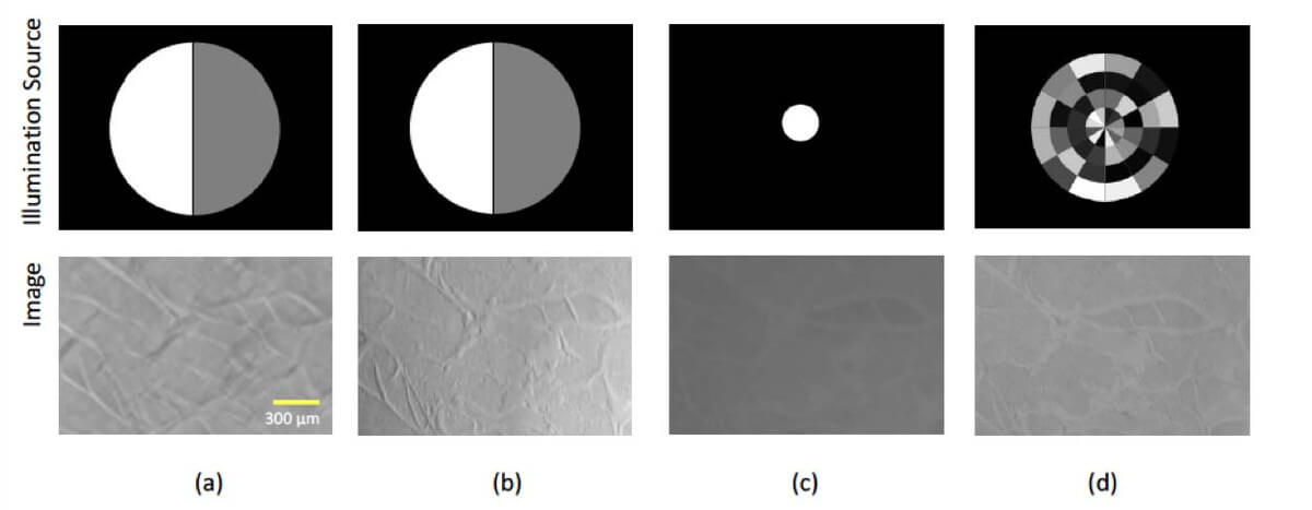
Results of the improved phase contrast using different illumination source shapes displayed on a homemade spatial light modulator.
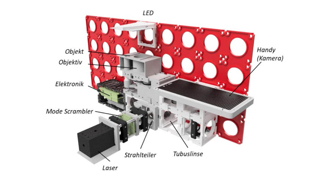
CAD drawing of the proposed microscope based on a modular framework.