- Home
- Research
- Fiber Research and Technology
- Research results
- UV spectroscopy in water-filled anti-resonant hollow core fibers
UV spectroscopy in water-filled anti-resonant hollow core fibers
28.03.2019
Monitoring water quality becomes increasingly important nowadays due to the augmented use of antibiotics worldwide. Residual amounts of many species can be quantified by UV spectroscopy due to strong absorption features in this regime. We show that water analysis inside an anti-resonant hollow core fiber potentially reaches higher concentration detection sensitivities than comparable methods.
By M. Nissen
Our society today faces various challenges, one of them: The contamination of our water supplies by pharmaceutical substances, such as antibiotics. To enable intervention at early stages, highly sensitive water monitoring methods become more and more important. One approach to quantify water-contaminating species is UV spectroscopy since biological substances often show strong absorption at wavelengths little below the visible regime. The sensitivity of current UV spectroscopic devices, however, is limited by relatively short light-matter interaction lengths, usually in the order of one centimeter, the typical light path length inside a measurement cuvette. The idea behind a new approach pursued at IPHT is to extend this interaction length to the order of meters by using liquid-filled hollow-core fibers – yielding a potential sensitivity increase up to two orders of magnitude.
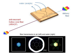
The fiber that is applied for the UV spectroscopic measurements is an anti-resonant hollow core fiber (ARHCF) fabricated at IPHT. It contains a hexagonally shaped core with a diameter of approximately 30 µm and guides light of wavelengths below 340 nm when filled with water. The comparably large channel diameter inside the fiber enables liquid filling or exchanging within seconds to minutes. At the same time, the required analyte volume is about three orders of magnitude smaller than that needed for typical cuvette based systems.
In order to simultaneously couple light into the ARHCF and fill it with analyte, the input section of the fiber is integrated into an optofluidic chip that was likewise fabricated at IPHT. The fiber channel inside the chip is accessible by the two opposite sides so that both the ARHCF and a delivery fiber can be threaded in and be brought closely together into a butt coupling position for direct light transmission. Thus the chip provides protection for the fiber-to-fiber junction against external influences and, even more important, allows to access the ARHCF with state-of-the-art microfluidics. Once the fiber-chip system is assembled, the fibers are fixed in their respective positions and their channel is sealed with UV curing glue, resulting in a monolithic system that can be introduced in a common spectroscopic setup by connecting a light source to the delivery fiber and placing a spectrometer behind the free output section of the ARHCF.
So far, the in-fiber UV spectroscopy has been applied for the concentration quantification of two different application-relevant pharmaceutical substances – sulfamethoxazole and sodium salicylate – that were dissolved in water. Concentration levels of the respective substances down to 0.1 µM (26 ppb) and 0.4 µM (64 ppb) could be measured. This is comparable to the performance of other methods, but can most likely be improved by further device integration, leading to less measurement-to-measurement variations and thus higher concentration detection accuracy. In any case, the results until now confirm that ARHCFs can indeed be applied in place of a measurement cuvette without complicating the measurement principle of common UV spectroscopy, and therefore open new perspectives for highly sensitive liquid-based analytics in fibers.
Funded by: DFG, Freistaat Thüringen and ESF