- Home
- Research
- Biophysical Imaging
- Research results
- New Microscopy Method sets Standards with Unrivaled Sharpness
New Microscopy Method sets Standards with Unrivaled Sharpness
07.12.2022
MINFLUX microscopy allows the smallest building blocks of life to be observed with unprecedented accuracy. Researchers at Leibniz IPHT are further developing super microscopy and experimenting with photostable fluorescence probes to unlock new potentials of this novel microscopy technique for applications in life sciences.
How do viruses infect cells and overcome the barrier of the cell membrane? How can pathogens multiply unhindered in the human body and spread to cause an infection? What happens inside the cell during an inflammation? How are signals transmitted at the cell envelope? Biology and medicine are trying to find answers to these and similar questions using microscopy. However, optical light microscopy quickly reaches its limits when observing the smallest details. Already in the 19th century, the physicist Ernst Abbe recognized that structures can only be precisely resolved microscopically down to a size of 200 nanometers. “Smaller details can only be perceived in a blur under the microscope and cannot be clearly distinguished optically,” explains Prof. Dr. Christian Eggeling, head of the Biophysical Imaging Department at Leibniz IPHT.
To overcome these physical limitations, researchers developed a super-resolution microscopy: Nobel prize winner and physicist Stefan Hell expanded the most established super-resolution microscopic methods STED and (f)PALM/(d)STORM to create the ultrahigh-resolution MINFLUX fluorescence microscopy. Instead of maximizing light intensities from the irradiated laser or detected fluorescent light, the MINFLUX approach minimizes light intensities. This enables razor-sharp insights into the inner of cells with a unique resolution of less than ten nanometers.
Observing three-dimensional insights into living cells with pin-sharp clarity
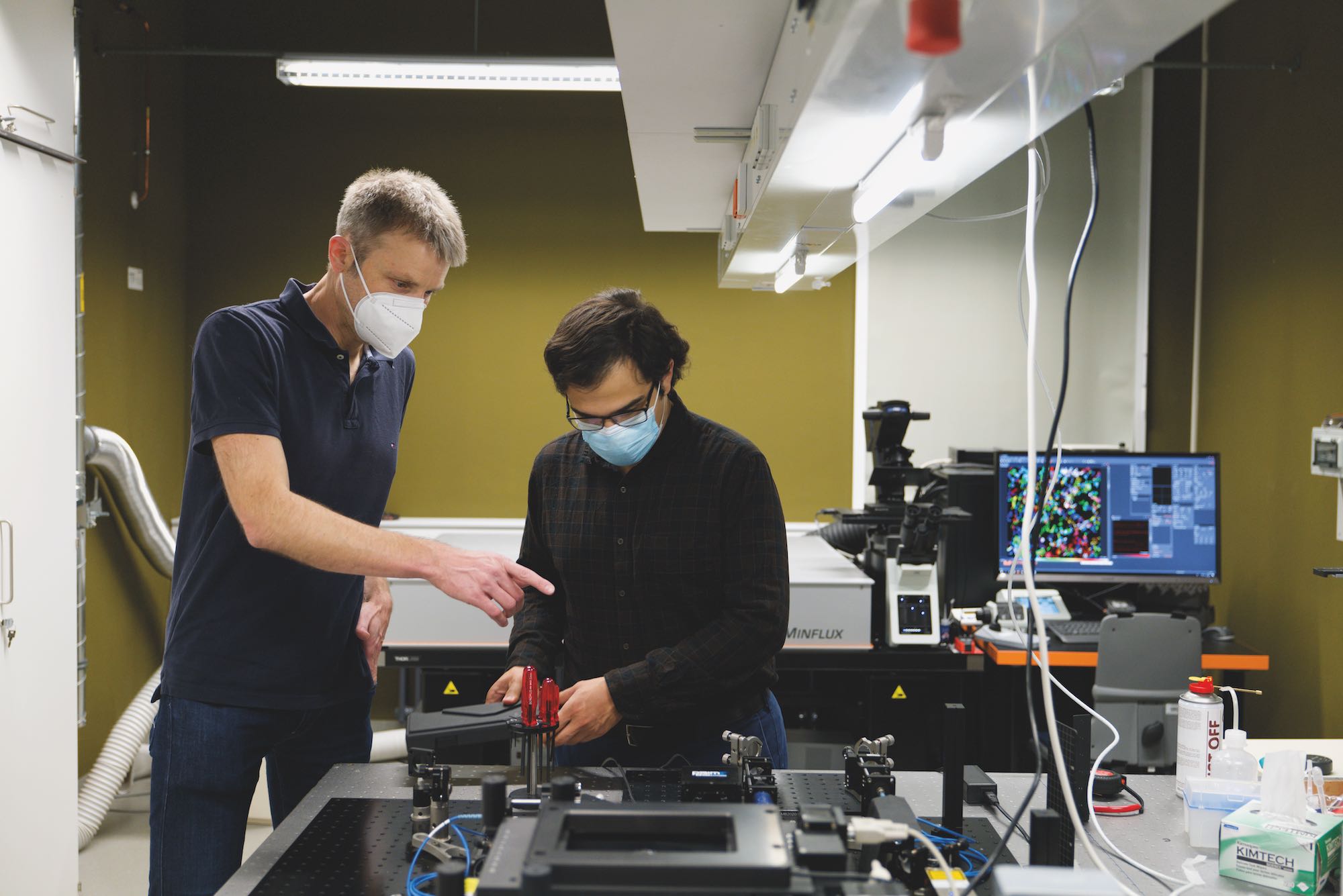
Scientists at Leibniz IPHT, among others, are researching the strengths of this supermicroscopy. In 2021, they received a MINFLUX device, which was acquired as part of the large-scale equipment initiative “Novel, Experimental Light Microscopes for Research” of the German Research Foundation.
“The MINFLUX technology allows completely new three-dimensional insights into living cells. With it, processes on the cell membrane can not only be made visible, it also significantly expands our understanding of the interplay of the cell components. Intracellular processes, such as the infection of cells with HIV or corona viruses, can thus be deciphered. In order to fully exploit the potential of MINFLUX microscopy, we are working on further technological developments, optimizing the hardware and methods of evaluation, for example, and studying its possibilities and limits. This paves the way to further improve the technology in order to enhance the support of biology and medicine in answering cell biological questions in the long term,” explains Christian Eggeling.
Color makes the difference
To track dynamics at the cell membrane microscopically, fluorescent dyes are needed. “With these dyes, we insert a label into the cells to be observed. These dyes glow thanks to the microscope’s excitation light. In this way, the movement of the molecules can be tracked in time and space, and their diffusion through the cell membrane can be precisely observed. Unfortunately, some of these dyes, the fluorophores, lose their ability to fluoresce in fractions of seconds due to photochemical processes, like a photograph whose colors fade,” says biophysicist Christian Eggeling. The fading of the dyes is a problem, especially in longterm observations.
Novel photostable dyes, also known as fluorescent probes, offer the potential to overcome the disadvantages of photobleaching and enable observations on cell membranes in real time with high temporal and spatial resolution as well as long acquisition times. In 2021, the team of Christian Eggeling and his colleague Pablo Carravilla tested new fluorescent probes based on Nile red, which were synthesized by their collaboration partner Prof. Dr. Andrey Klymchenko from the Université de Strasbourg. These markers only accumulate in the cell membrane for a short time, and only there they begin to fluoresce when stimulated by light irradiation.
Fluorescence probes can be used, for example, to observe the fusion of membrane vesicles with the cell membrane, the so-called membrane fusion, as well as the union of lipid layers. The new dyes thus provide the opportunity to gain detailed insights into the molecular mobil
ity on biological membranes via high-resolution time-lapse imaging or real-time imaging. Since MINFLUX technology requires low concentrations of fluorescent probe for its high precision, these dyes that only accumulate for short times would also be suitable for this technology.
“In the future, we want to further investigate the performance of super-resolution STED and MINFLUX microscopy techniques to make them useful for biomedical applications. The technology has the potential to inspire pharmaceutical research and modern medicine,” says Christian Eggeling full of hope.
More information in teh following viedo:
Unfortunately, this video can only be downloaded if you agree to the use of third-party cookies & scripts.