Laser light detects tumors: How novel optical imaging strategies help to improve cancer diagnostics

4. Februar 2021
A new book provides in-depth description of state-of-the-art optical approaches for the enhancement of earlier cancer detection and classification
Cancer — this diagnosis is hitting more and more people in our aging society. But the earlier the disease is detected, the greater the likelihood that it can be treated effectively. Modern light-based technologies can help to decisively improve the diagnosis and treatment of cancer, because they can produce diagnostically relevant information quickly, reliably and gently.
Minimally invasive examination for accurate diagnosis
The recently published book on “Multimodal Optical Diagnostics of Cancer” by the international biophotonics experts Jürgen Popp, Valery V. Tuchin and Valery Zakharov now provides an in-depth description and discussion of the latest optical methods for non-invasive cancer diagnostics and treatment. Researchers in biomedical engineering, photonics and medicine will find the book to be a guide to optical approaches for cancer diagnostics and screening, for long-term monitoring to image-guided intervention.
“Light-based methods are a promising tool to facilitate early detection and diagnosis of cancer,” says Prof. Jürgen Popp, scientific director of the Leibniz Institute of Photonic Technology (Leibniz IPHT) in Jena. He co-edited the volume with Prof. Valery V. Tuchin of Saratov State University and Prof. Valery Zakharov of the National Research University in Samara, Russia.
“The multimodal approach provides a wealth of information: for example, on the structure and morphology of the tissue and its molecular composition. This overall picture helps medical professionals make an accurate diagnosis and choose the appropriate treatment. For patients, in turn, modern optical methods can make the examination easier because they are non-invasive or minimally invasive and label-free.”
The vision: cancer operations without a scalpel
One example is the diagnosis of tumor tissue: the current gold standard is examination by the trained eye of physicians followed by removal of a tissue sample for analysis under the microscope. In this way, patients only find out after up to four weeks whether the entire tumor has really been removed during a cancer operation. Multimodal imaging, on the other hand, makes cancerous tissue directly visible with laser light. This could help surgeons to remove tumors more precisely — and in the future even make cancer operations possible without a scalpel.
„The appearance of this book was stimulated by the recent rapid progress in novel photonics technologies,” explains Jürgen Popp. “These, in turn, are spurring research into new diagnostic procedures.” For example, novel fiber-optic instruments could open a path to minimally invasive medicine. “Flexible endoscopes can not only identify the tumor, but remove it right away: ablate it layer by layer based on light.”
Thus, in addition to outlining optical imaging strategies, the editors provide a glimpse of how novel methods for noninvasive diagnostics may pave the way to new diagnostics and therapies for cancer.
//
Jürgen Popp, Valery V. Tuchin, Valery Zakharov (Hg.): Multimodal Optical Diagnostics of Cancer. Springer, Heidelberg 2020.
ISBN 978-3-030-44593-5
Jürgen Popp, Valery V. Tuchin, Valery Zakharov (Hg.): Multimodal Optical Diagnostics of Cancer. Springer, Heidelberg 2020.ISBN 978-3-030-44593-5
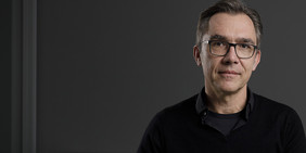
Co-editor Prof. Jürgen Popp, scientific director of Leibniz IPHTPhoto: Sven Döring/ Leibniz-IPHT
Unfortunately, this video can only be downloaded if you agree to the use of third-party cookies & scripts.
Contact
Related News

Third party cookies & scripts
This site uses cookies. For optimal performance, smooth social media and promotional use, it is recommended that you agree to third party cookies and scripts. This may involve sharing information about your use of the third-party social media, advertising and analytics website.
For more information, see privacy policy and imprint.
Which cookies & scripts and the associated processing of your personal data do you agree with?
You can change your preferences anytime by visiting privacy policy.