Hazel, birch or grass?

27. Juli 2018
Distinguish pollen using microfluidics and neuronal networks
A miniaturized lab on a chip enables high resolution microscopy images of several thousands of pollen particles in seconds. Neuronal networks take over the image processing and classify the particles fast and reliable. Andreas Kleiber, PhD student at the Leibniz-Institute of Photonic Technology in Jena (Leibniz IPHT), tested the method on several highly allergenic pollen. For his results, presented during the 3rd Imaging Technology Summer Workshop dedicated to Big Data in Imaging, Kleiber was awarded the poster price by the European Society for Molecular Imaging.
Up to 1000 pollen per second flow by the optical window in a narrow channel on the stamp sized chip. A digital camera captures each of the tiny single grains through a microscope lens. To receive sharp shots for the following data processing, every analyzed particle has to pass the liquid channel in the focal plane of the lens. The height of the focal plane of the used high-resolution lenses measures less than a hundredth millimetre.
Scientists of Leibniz IPHT met this technological challenge employing a sophisticated design of the components in the microfluidic chip. The patented method enables them to align the pollen grains exactly in the focal plane and therefore to obtain sharp images of all objects. “Using two liquid streams from the sides, we press the particle stream to a sheet, just like a nozzle. A new arrangement of the micro channels rotates the sheet by 90° into the focal plane,“ explains Andreas Kleiber the technology. in the scope of his PhD thesis, the scientist researches methods for the high-throughput analysis of bioparticles using microfluidic chips.
The principle of hydrodynamic focusing is already known in the field of flow cytometry for the analysis of cell populations. Here, the cells are focused in a way that they pass by the measurement window along a line. “New to our system is, that we arrange the particles in a thin, two dimensional lamella, and therefore use the whole frame of the camera. This makes the method so rapid“, says Kleiber. The researchers can actuate the horizontal position and thickness of the particle layer accurately. Therefore, they are able to control the rotation of the pollen in the stream. “Using methods already known from computer-tomography we are able to produce 3D-image data that contain important information e.g. about the three-dimensional morphology of a pollen grain. The 3D-information improves the reliability of the pollen identification significantly“, elucidated Kleiber. The researcher evaluates images of the different pollen with software tools for particle tracking and feature extraction. A pre-trained convolutional neuronal network classifies the shots to a certain kind of pollen by means of the extracted data. The hit rate is above 98%.
The researchers classified the pollen, which originate from the research group Indoor Climatology at the University Hospital Jena, without any additional label, solely on basis of the image information from microscopy. „We are able to use the method furthermore for the analysis of cells e.g. to distinguish subtypes of white blood cells“, underlines Dr. Thomas Henkel, who leads the relevant research work at Leibniz IPHT. “In the future, it should be possible to sort bioparticles with our chip“, says Henkel about the planned research, which is funded by the EU in the range of the Era-NET-DLR project “WaterChip“.
Birch pollen is one of eight different types of pollen, investigated by Andreas Kleiber.Picture: designed by Bearfotos – Freepik.com
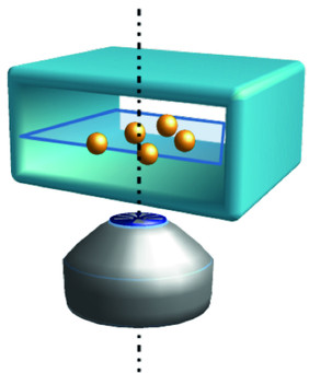
The patented design of the microfluidic channels allows to align all particles in the focal plane. Source: A. Kleiber/Leibniz-IPHT
Contact
Related News

Third party cookies & scripts
This site uses cookies. For optimal performance, smooth social media and promotional use, it is recommended that you agree to third party cookies and scripts. This may involve sharing information about your use of the third-party social media, advertising and analytics website.
For more information, see privacy policy and imprint.
Which cookies & scripts and the associated processing of your personal data do you agree with?
You can change your preferences anytime by visiting privacy policy.