- Home
- Research
- Spectroscopy and Imaging
- Research results
- Metabolite Detection Using SERS
Metabolite Detection Using SERS
14.07.2020
To make specific detection of metabolites possible in medically relevant concentrations, surface-enhanced Raman spectroscopy (SERS) is applied because it allows for molecularly specific and highly sensitive detection images. We achieved the detection of two relevant metabolites (i.e., pyocyanin and pyrazinoic acid) using SERS-based detection images.
By Olga Žukovskaja //Izabella J. Jahn // Anna Muehlig // Karina Weber // Dana Cialla-May // Juergen Popp
In order to take measures quickly in terms of the correct treatment of infectious diseases, both the fast identification of the pathogens and information regarding drug resistance are required. Surface-enhanced Raman spectroscopy (SERS) is an attractive tool to address this analytical issue. We illustrated the potential of the SERS technique in bioanalytics via the characterization and investigation of two metabolites (i.e., pyocyanin and pyrazinoic acid) in artificial matrices with different complexity.
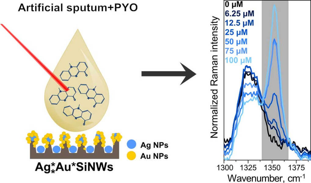
Pyocyanin (PYO) is a specific metabolite of the Pseudomonas aeruginosa bacteria, and its detection directly in sputum samples can significantly reduce the diagnosis time of respiratory infections. Therefore, a sensitive, rapid, easy-to-use, and cost-effective PYO detection platform is needed. In our study, SERS was suggested for this clinical task. Keeping in mind the requirement of a simple and cheap detection platform, silicon nanowires (SiNWs) that are produced chemically within 20 min. and variously decorated with silver and/or gold nanoparticles were introduced as SERS substrates. By investigating different designs, it was concluded that silver nanoparticles on the bottom of the nanowires is beneficial for an intensive SERS signal. Moreover, placing bimetallic silver/gold nanoparticles on the top of SiNWs rather than just gold nanoparticles improved the stability of the signal. Using such substrates, the detection of PYO was performed in artificial sputum, which mimics clinical samples. To decrease the influence of the matrix, a dilution of the samples was suggested and a SERS PYO signal at a concentration of as low as 6.25 µM was able to be observed (see Figure 1). Moreover, a linear response of the SERS intensity in the clinically relevant concentration range of 6.25-100 µM was achieved. These results indicate that label-free SERS is a suitable candidate for PYO detection in respiratory bodily fluids. This technique holds significant promise for quantitative analysis.
As a further example, the treatment of tuberculosis (TB) faces challenges in many developing countries due to the presence of multidrug resistant pathogens. Pyrazinamide (PZA) is one of the first line agents administered for drug-susceptible TB cases. Nevertheless, PZA susceptibility testing is challenging. PZA is a prodrug; only its metabolite, pyrazinoic acid (POA), can kill bacteria. PZA resistance is associated with a very low PZA-to-POA conversion or none at all. PZA resistance could, therefore, be assessed by quantifying POA in the extracellular environment of an in vitro liquid culture. POA and PZA show distinct SERS signatures, making the quantification of both substances possible in a mixture. To assure reproducible measurement conditions, we used a droplet-based microfluidic platform with in-situ synthesized Ag nanoparticles. A linear dynamic range from 20 µM to 100 µM for PZA and 50 µM to 100 µM for POA was found. The ratio of the marker modes of PZA and POA can be a relevant indicator of PZA resistance.
In summary, we showed the potential of the SERS technique in the detection of metabolites in medically relevant concentrations. In future work, the transfer of these detection plans to real samples is envisaged.
Funded by BMBF, DLR