Cancer Surgery: Jena Research Team Develops Imaging Fiber Endoscope for Tissue Diagnostics
14. Oktober 2021
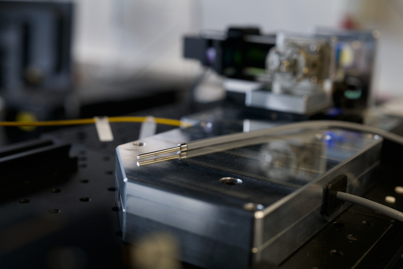
With current methods, only after a cancer operation it is possible to determine with certainty whether the entire tumor has actually been removed. An interdisciplinary research team from Jena has now presented a new type of fiber endoscope that could be used in the future to visualize tumor margins directly inside the body during surgery. The probe is based on a specially developed multimohedral fiber and provides tissue images that contain both morphological and biochemical information. The researchers from Leibniz Institute of Photonic Technologies, Friedrich Schiller University Jena and the company GRINTECH published their results on October 5, 2021 in the renowned journal “Light: Science & Applications” (DOI 10.1038/s41377-021-00648-w).
It can take several days for patients to be certain that a cancer operation was successful. Only the subsequent histopathological examination of a biopsy taken provides certainty as to whether the entire tumor has actually been removed. The fiber endoscope developed by the Jena research team, on the other hand, opens up the possibility of achieving a diagnosis in real time. The probe combines three imaging techniques at once and delivers high-resolution tissue images from inside the body. They contain both morphological and biochemical information.
Diagnosis in real time instead of after several days
“The endoscope offers the potential to quickly and reliably distinguish between healthy and diseased tissue – and to do so in vivo, i.e. in a minimally invasive examination in which the probe is placed directly on the suspicious tissue,” explains Prof. Jürgen Popp, scientific director of the Leibniz Institute of Photonic Technologies (Leibniz IPHT) in Jena, under whose leadership the novel probe was researched.
Flexible probe for minimally invasive examinations
For this purpose, the fiber technology team at Leibniz IPHT developed a special microstructured optical fiber. In combination with an intelligent and ultra-compact optical concept from the company GRINTECH, it leads to a completely fiber-based endoscopic setup for multimodal nonlinear endoscopy. It captures tissue images as currently done with a commercially available, bulky laser scanning microscope, while being comparatively inexpensive to manufacture. “In the future, the novel multimodal image probe could open up new possibilities for label-free tissue diagnostics in surgery and endoscopy – for example, to clearly identify tumor margins during surgery,” says Jürgen Popp.
More accurate detection of tumor margins
This would not only help to improve the chances of cure, but could also save considerable costs in the healthcare system by helping to avoid expensive and stressful follow-up treatments for patients. Currently, in the case of tumors in the head and neck area, for example, cancer cells are subsequently found after almost every 10th operation.
Success story from the Jena optics site
The technological realization of the patented imaging fiber optics is the result of many years of cooperation between the Jena researchers and the micro-optics specialist GRINTECH. “Our know-how in the field of endomicroscopic probes, which we have now extended to the use of miniaturized scanners with corresponding control and image processing software, and the expertise of Leibniz IPHT in the development of microstructured glass fibers have ideally complemented each other,” says Dr. Bernhard Messerschmidt of GRINTECH, which was founded at the end of 1999 as a spin-off of the Jena-based Fraunhofer Institute for Applied Optics and Precision Engineering. “In this respect, this joint development is also a success story of Jena as an optics and photonics location – both in the close networking of science and industry and in our collaboration in highly efficient interdisciplinary teams here,” adds Jürgen Popp.
Award-winning method for rapid cancer diagnostics
The joint development of the endoscopy probe for biomedical imaging was funded by the German Federal Ministry of Education and Research (BMBF) from 2017 to 2019 as part of the Regional Growth Core “Tailored Optical Fibers – TOF”. The optical fast method for multimodal tissue diagnostics with AI-supported evaluation is based on a method developed by a team from Leibniz IPHT, Friedrich Schiller University and Jena University Hospital, and the Fraunhof Institute of Applied Optics and Precision Engineering (IOF). For this optical approach to rapid cancer diagnostics, the interdisciplinary Jena team was awarded the prestigious Kaiser Friedrich Research Prize in 2018. The underlying research project entitled “CDIS Jena – Cancer Diagnostic Imaging Solution Jena” was funded by BMBF, by the German Research Foundation (DFG) and by the Thuringian Ministry of Economy, Science and Digital Society (TMWWDG).
Publication:
Ekaterina Pshenay-Severin, Hyeonsoo Bae, Karl Reichwald, Gregor Matz, Jörg Bierlich, Jens Kobelke, Adrian Lorenz, Anka Schwuchow, Tobias Meyer-Zedler, Michael Schmitt, Bernhard Messerschmidt, Juergen Popp: Multimodal nonlinear endomicroscopic imaging probe using a double-core double-clad fiber and focus-combining micro-optical concept, Light: Science & Applications (2021) 10:207 https://doi.org/10.1038/s41377-021-00648-w
Contact

Scientific Contact

Prof. Dr. Jürgen Popp