- Home
- Research
- Spectroscopy and Imaging
- Work-Groups
- Raman and IR spectroscopic analysis
Raman and IR spectroscopic analysis
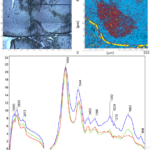
The work group researches the application of Raman and infrared based spectroscopy and imaging techniques to characterize cells and tissues as well as to detect low molecular weight biomolecules such as drugs or metabolites. To characterize cells and tissues, Raman spectrometer systems are coupled with microscopes, fiber-optic probes, or microfluidic chips, and the vibrational spectroscopic data obtained are analyzed using machine learning methods. Validation is performed using histopathological examinations as the gold standard.
Absorption of radiation in the mid-infrared range between 2.5 and 11 µm is also used to measure a complementary vibrational spectroscopic fingerprint. In addition to classical Fourier transform infrared spectrometers, novel laser direct infrared (LDIR) and optical photothermal infrared (O-PTIR) techniques with tunable quantum cascade lasers are also applied.
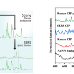
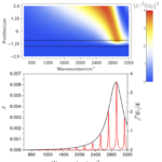
In addition, the activities of the group include the study of optical effects to increase the signal intensity in vibrational spectroscopy by several orders of magnitude. This enhancement is based on the amplification of the electric field intensity near photonic and plasmonic nanostructures and on a coupling of their modes with the molecular vibrations. Based on a fundamental understanding of this signal amplification, goals are to design, investigate and apply appropriate structures. The applications fields are in the area of environmental analysis as well as in the medical sector, such as the monitoring of drug levels and the detection of metabolites and pathogens. In line with the Leibniz IPHT motto Photonics for Life, the group's goal is to work with medical professionals to transfer Raman- and infrared-based methods into the clinic for improved healthcare.
Research Insights
Unfortunately, this video can only be downloaded if you agree to the use of third-party cookies & scripts.
Research Topics
Many years of experience in sample preparation and the instrumental infrastructure are available in the research group to perform bioanalytical investigations on cell and tissue samples. Calibration procedures of the instruments allow to record series of measurements under reproducible conditions. Furthermore, the group works on understanding, designing and engineering photonic and plasmonic nanostructures including functionalization to amplify vibrational bands in spectra and apply high performance SERS and SEIRA substrates for biophotonics.
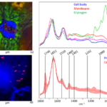
- Raman microscopic imaging at subcellular resolution
- Raman spectroscopy with fiber optic probes
- Optical photothermal infrared spectroscopy and imaging with quantum cascade laser and optional Raman spectroscopy and fluorescence microscopy
- Fourier transform infrared imaging in transmission and reflection with focal plane array detector
- Laser Direct Infrared Spectroscopy and Imaging with Quantum Cascade Laser
- Understanding spectral intensities in IR and VCD spectra
- Exploration of photonic and plasmonic nanostructures for use as SERS or SEIRA substrates, respectively
- Application of the SERS or SEIRA method in biological, environmental and medical analytics
- Detection of drugs and their metabolites and their quantification in complex body fluids such as urine or blood-based matrices or biological samples
- Analysis of body fluids for the detection of biomarkers
- Chiral substrates for surface-enhanced enantiomer identification
- Surface functionalization of plasmonic nanostructures
Areas of application

- Innovative single cell diagnostics as liquid biopsy to identify tumor cells circulating in body fluids of cancer patients
- Raman and infrared imaging of thin sections as a marker-free, molecular contrast technique to distinguish pathological from control tissues
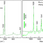
- Identification of microplastics using simultaneous infrared and Raman spectroscopy
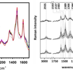
- Quantitative detection of low molecular weight substances (e.g. drugs, metabolites) in body fluids and biological samples using powerful SERS-active structures
- Identification of biomarkers for therapy-supporting investigation of body fluids
- Rapid detection of pathogenic agents
- Simplified and more sensitive identification of enantiomers
- SERS-based detection methods in clinical settings and using resource-efficient SERS structures for applications with low laboratory/clean room infrastructure


