Laser Light Detects Tumors: Jena Researchers Present Device for Rapid Tissue Analysis
21. Juni 2019
Cancer – this diagnosis affects almost every second German at some point in his life. It is the second most frequent cause of death in Germany. But the earlier the disease is diagnosed, the greater are the chances of surviving it. A team of researchers from Jena will present a groundbreaking new method for the rapid, gentle and reliable detection of tumors with laser light at the leading trade fair „Laser World of Photonics“ from 24 to 27 June 2019 in Munich. For the first time, the Leibniz Institute of Photonic Technology (Leibniz IPHT) will present a compact device for rapid cancer diagnosis during surgery. The optical method will help surgeons to remove tumors more precisely and could make cancer operations possible without a scalpel.
It can take up to four weeks before patients can be sure whether the entire tumor has been removed during cancer surgery. A time of agonizing uncertainty — in which any remaining tumor cells can already multiply again. A team of scientists from Jena has now researched a diagnostic procedure that could revolutionize the previous procedure: Using laser light, the researchers make cancerous tissue visible. This enables them to provide the surgical team with real-time information in order to reliably identify tumors and tumor margins and decide how much tissue needs to be cut away.
This is made possible by a compact microscope developed by a research team from the Leibniz Institute of Photonic Technology, Friedrich Schiller University, the University Hospital and the Fraunhofer Institute for Applied Optics and Precision Engineering in Jena. It combines three imaging techniques and uses tissue samples to generate spatially high-resolution images of the tissue structure during surgery. Software makes patterns and molecular details visible and processes them with the aid of artificial intelligence. The automated analysis is faster and promises more reliable results than the currently used frozen section diagnostics, which can only be evaluated by an experienced pathologist and still have to be confirmed afterwards.
The optical method, for which the Jena scientists were awarded the renowned Kaiser Friedrich Prize in 2018, helps to prevent weakened patients from having to undergo another operation. It thus makes a significant contribution to improving their chances of recovery. Professor Jürgen Popp, scientific director of Leibniz IPHT, who was also involved in researching the laser rapid test, predicts that the compact microscope could be in the clinic in five years‘ time.
This could save the German healthcare system considerable costs. „One minute in the operating room is the most expensive minute in the entire clinic,“ explains Professor Orlando Guntinas-Lichius, Director of the Department of Otolaryngology at the University Hospital Jena. In the case of tumors in the head and neck area, for example, cancer cells are found after almost every 10th operation.
And the Jena researchers are already thinking ahead. They are researching a solution that would enable them to use the unique properties of light to detect tumors inside the body at an early stage and remove them immediately. „To do this, we need novel methods that no longer work with rigid optics, but with flexible endoscopes,“ says Jürgen Popp. Technologists at Leibniz IPHT produce such fiber probes: glass fibers that are thinner than a human hair. They open the way to minimally invasive medicine that makes gentle diagnosis and healing possible. „Our vision,“ says Jürgen Popp, „is to use light not only to identify the tumor, but to remove it immediately. This would eliminate the need for physicians to cut with a scalpel and would enable them to ablate the tumor layer by layer using light in order to remove the tumor from the patient completely“. In ten to fifteen years, the research team hopes to find a solution. Popp predicts that this would „be a giant step towards completely new tumor diagnostics and therapy“.
The research work was funded by the European Union, the Federal Ministry of Education and Research, the Free State of Thuringia, the German Research Foundation, the Foundation for Technology, Innovation and Research Thuringia and the Chemical Industry Fund.
Exhibition stand at the „Laser World of Photonics“
Leibniz-IPHT will present the new device for cancer diagnosis at the exhibition stand „Photonics in the life sciences“ in Hall B2.350.
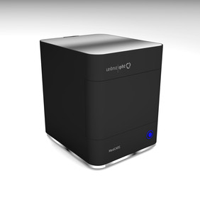
For routine applications in biomedicine and hospitals, the Jena research team integrated the multimodal imaging approach into an easy-to-use, multi-contrast nonlinear microscope (MediCARS). Picture: Leibniz-IPHT
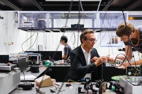
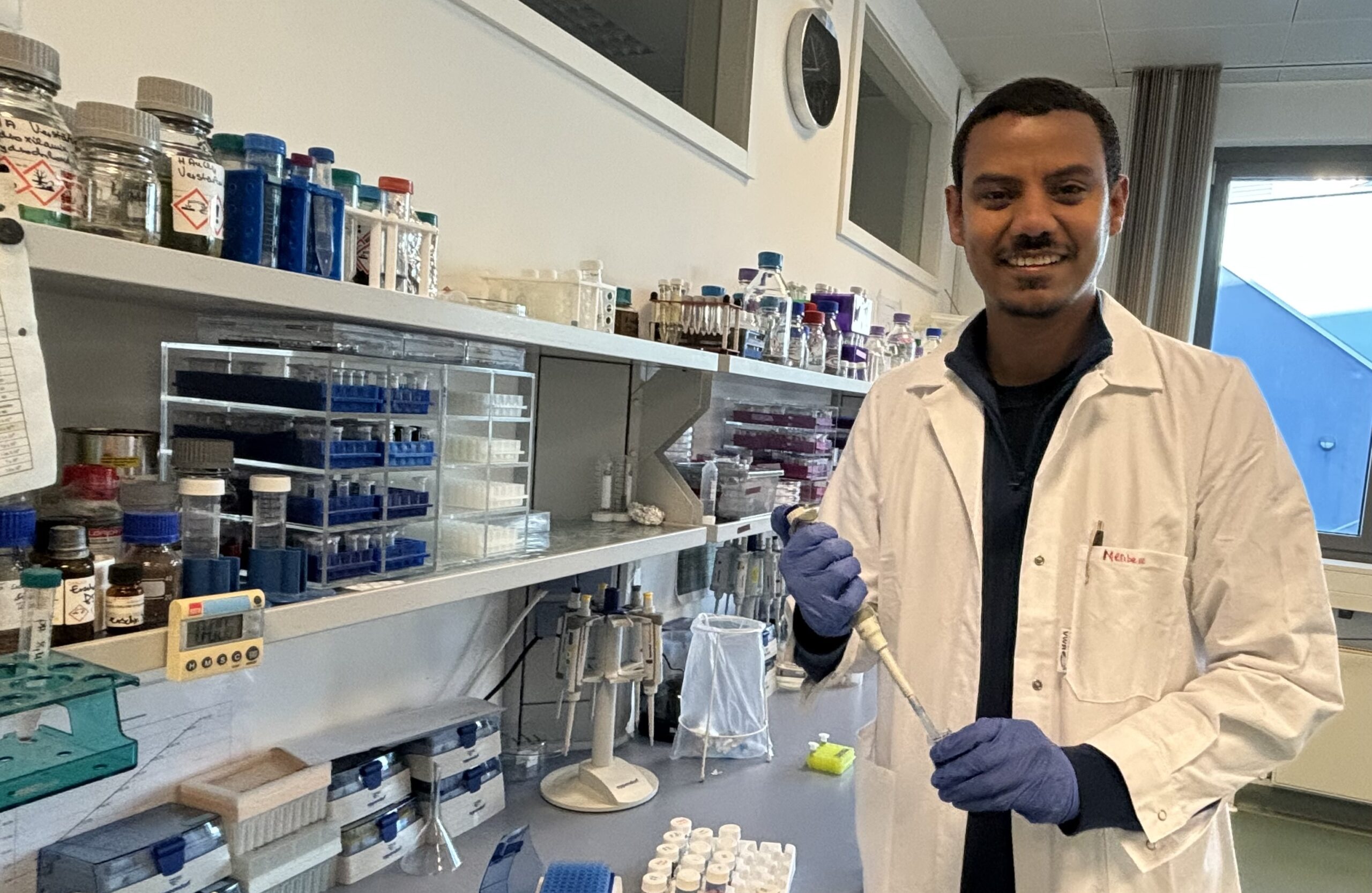
Jürgen Popp and Tobias Meyer researched the rapid method for cancer diagnostics together with a team of scientists from Jena. Photo: Sven Döring/ Leibniz-IPHT
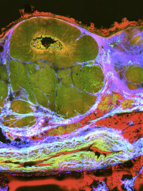
Multi-contrast image of a tissue affected by skin cancer. Using three different optical-spectroscopic techniques, morphological and molecular aspects become visible, enabling the surgical team to reliably identify cancerous tissue. Source: Leibniz-IPHT
Dieses Video kann leider nur geladen werden, wenn Sie der Verwendung von Drittanbieter Cookies & Skripten zustimmen.
Kontakt

Ähnliche Meldungen
Cookies & Skripte von Drittanbietern
Diese Website verwendet Cookies. Für eine optimale Performance, eine reibungslose Verwendung sozialer Medien und aus Werbezwecken empfiehlt es sich, der Verwendung von Cookies & Skripten durch Drittanbieter zuzustimmen. Dafür werden möglicherweise Informationen zu Ihrer Verwendung der Website von Drittanbietern für soziale Medien, Werbung und Analysen weitergegeben.
Weitere Informationen finden Sie unter Datenschutz und im Impressum.
Welchen Cookies & Skripten und der damit verbundenen Verarbeitung Ihrer persönlichen Daten stimmen Sie zu?
Sie können Ihre Einstellungen jederzeit unter Datenschutz ändern.